Dr. Akshay Shetty Orthopaedics
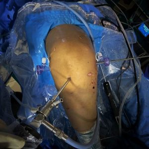
Knee Arthroscopy
Knee Arthroscopy
ACL Reconstruction
Anterior cruciate ligament known as ACL, located in the centre of the knee, that connects the bottom of the bone (femur) & top of the shin box(tibia). ACL is one of the majors stabilizes of the knee joint tear ruptures of the ligament resulting in knee instability.
PCL Reconstruction
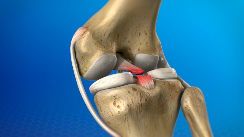
The posterior cruciate ligament (PCL) is located in the knee, just behind the anterior cruciate ligament (ACL). It is one of the ligaments that connects the shinbone (tibia) to the thighbone (femur). The posterior cruciate ligament restricts the tibia from moving backward with respect to the femur.

Meniscus Tear
A meniscus in the knee is a rubbery C-shaped structure that cushions the adjacent surfaces of the shin bone (tibia) and thigh bone (femur) at the level of the knee joint. Meniscus acts like shock absorbers.

MPFL Reconstruction
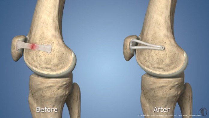
The Medial Patello- Femoral ligament (MPFL) is a part of the complex network of soft tissues that stabilize the knee. The MPFL is a ligament that stabilizes patella (kneecap) to the thigh bone (Femur) .

MCL / LCL Reconstruction
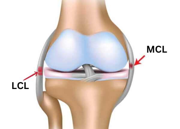
The MCL (Medial Collateral Ligament) and LCL (Lateral Collateral Ligament) are bands of tissue that connect the thigh bone to the shin bone at the level of the knee joint on either side and help stabilise the knee. The MCL is on the inner side of the knee, while the LCL is on the outer side of the knee
Cartilage Procedures
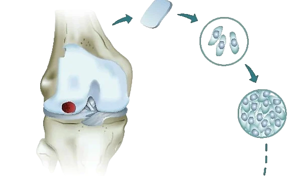
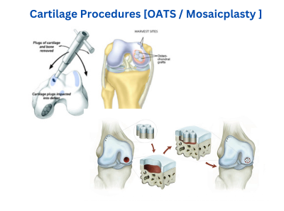
Articular cartilage is a firm, rubbery cushion-like material that covers the ends of bones in the knee joint. It prevents friction and rubbing of adjacent bones against each other in the joint and acts as a “shock absorber.”

Autologous Chondrocyte
Autologous Chondrocyte Implantation (ACI) is a process whereby articular cartilage cells, known as chondrocytes, are collected from patients.
